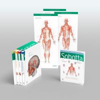
Sobotta Atlas of Anatomy, Package, 17th ed., English/Latin
General Anatomy and Musculoskeletal System; Internal Organs; Head, Neck and Neuroanatomy; Muscles Tables; Poster Collection
MORE THAN AN ATLAS
Studying anatomy is fun! Recognising the structures on the dissection, understanding their relationships and gaining
an overview of how they work together assures confident study and transition into clinical practice.
The Sobotta Atlas shows authentic illustrations of the highest quality, drawn from genuine specimens, guaranteeing
the best preparation for the gross anatomy class and attestation.
Sobotta focuses on the basics, making it totally comprehensive. Every tiny structure has been addressed according to
current scientific knowledge and can be found in this atlas. Themes relevant to exams and sample questions from oral
anatomy exams help to focus the study process.
The Sobotta Atlas is the optimal learning atlas for studying, from the first semester till the clinical semester. Case studies
present examples and teach clinical understanding. Clinical themes and digressions into functional anatomy are motivating
and impart valuable information for prospective medical practice.
With over 100 years of experience in 17 editions and thousands of unique anatomical illustrations, Sobotta achieves
ongoing success.
The volume General Anatomy and Muscoloskeletal System contains the chapters:
General Anatomy
Anatomical planes and positions - Surface anatomy - Development - Musculoskeletal system - Neurovascular pathways - Imaging methods - Skin and its derivatives
Trunk
Surface - Development - Skeleton - Imaging methods - Musculature - Neurovascular pathways - Topography, dorsal trunk wall - Female breast - Topography, ventral trunk wall
Upper Limb
Surface - Development - Skeleton - Musculature - Neurovascular pathways - Topography - Cross-sectional images
Lower Limb
Surface - Skeleton - Musculature - Neurovascular pathways - Topography - Cross-sectional images
The volume Inner Organs contains the chapters:
Organs of the thoracic cavity
Topography - Heart - Lung - Oesophagus - Cross-sectional images
Organs of the abdominal cavity
Development - Topography - Stomach - Intestines - Liver and gallbladder Pancreas - Neurovascular pathways - Cross-sectional images
Retroperitoneal space and pelvic cavity
Topography - Kidney and adrenal gland - Efferent urinary tracts - Rectum and anal canal - Male genitalia - Female genitalia - Cross-sectional images
The volume Head, Neck and Neuroanatomy contains the chapters:
Head
Overview - Skeleton and joints - Adipose tissue and scalp - Musculture ?? Topography - Neurovascular pathways - Nose - Mouth and oral cavity - Salivary glands
Eye
Development - Skeleton - Eyelids - Lacrimal gland and lacrimal apparatus - Muscles of the eye - Topography - Eyeball - Visual pathway
Ear
Overview - Outer ear - Middle ear - Auditory tube - Inner ear - Hearing and equilibrium
Neck
Overview - Musculature - Pharynx - Larynx - Thyroid gland - Topography
Brain and spinal cord
Development - General principles - Brain ?? Meninges and blood supply - Cerebral areas - Cranial nerves - Spinal cord - Sections
Sobotta – learning tables for muscles, joints and nerves
The most important facts about muscles, joints and nerves are summarised in 62 tables, helping you to revise
methodically and efficiently!
- The origin, insertion, innervation and function of all muscles in the human body are presented in tabular form.
The relevant muscle is shown in situ each time. - The tables for branches and innervation areas of the Plexus cervicalis, brachialis and lumbosacralis give
a good overview for a clinical diagnosis. - The most important joints of the upper and lower limbs are shown in tables and additionally in schematic
illustrations. - The tables for neuroanatomy give the cranial nerves, cortical areas and nuclei of the thalamus.
Tables for the cranial nerves identify and compare the pathways, supply areas and fibre qualities.
Compatible to the 17th edition of the Sobotta Atlas!
Herausgeber*innen / Autor*innen
Prof. Dr. Friedrich Paulsen
Friedrich Paulsen wurde 1965 in Kiel geboren und absolvierte nach dem Abitur in Braunschweig zunächst eine Ausbildung als Krankenpfleger. Anschließend studierte er Humanmedizin an der Christian-Albrechts-Universität zu Kiel (CAU). Nach dem AiP an der Uniklinik für Mund-, Kiefer- und Gesichtschirurgie und einer Zeit als Assistenzarzt in der Universitäts-HNO-Klinik wechselte er 1998 an das Anatomische Institut der CAU, wo er 1997 seine Promotion abgeschlossen hatte und sich 2001 für das Fach Anatomie habilitierte.
2003 erhielt er Rufe auf C3-Professuren für Anatomie an die LMU München und die MLU Halle-Wittenberg. In Halle gründete er ein Weiterbildungszentrum für Klinische Anatomie. Nach einem weiteren abgelehnten Ruf an die Universität des Saarlandes ist er seit 2010 Professor für Anatomie und Institutsdirektor an der Friedrich-Alexander-Universität Erlangen-Nürnberg (FAU). Hier lehnte er weitere zwischenzeitlich ergangene Rufe renommierter Universitäten ab.
Friedrich Paulsen ist Ehrenmitglied der Anatomical Society (Great Britain and Ireland) und mit zahlreichen wissenschaftlichen Preisen ausgezeichnet, unter anderem dem Dr. Gerhard Mann SICCA-Forschungspreis, dem SICCA-Forschungspreis des Berufsverbands der Augenärzte Deutschlands und der Commemorative Medal der Comenius-Universität Bratislava. Darüber hinaus erhielt er mehrere Lehrpreise.
Seine Forschungsschwerpunkte liegen auf dem Gebiet der angeborenen Immunabwehr der Augenoberfläche und der Erforschung der Ursachen des trockenen Auges. Forschungsaufenthalte führten ihn nach Spanien und Großbritannien. Er ist Herausgeber der Zeitschrift Annals of Anatomy.
Prof. Dr. Jens Waschke
Jens Waschke (geb. 1974 in Bayreuth) hat an der Universität Würzburg Medizin studiert und 2000 bei Prof. Dr. med. Detlev Drenckhahn in der Anatomie promoviert. Nach dem AiP in der Anatomie und Inneren Medizin habilitierte er sich 2007 für das Fach Anatomie und Zellbiologie. Zwischen 2003 und 2004 verbrachte er einen neunmonatigen Forschungsaufenthalt an der University of California in Davis bei Professor Fitz-Roy Curry. Von 2008 an war er Inhaber des neu gegründeten Lehrstuhls III an der Universität Würzburg, bevor er an die Ludwig-Maximilians-Universität in München berufen wurde. Dort leitet er seit 2011 an der Anatomischen Anstalt den Lehrstuhl I für Vegetative Anatomie. Jens Waschke engagiert sich in der Anatomischen Gesellschaft als Prüfer für den Fachanatom sowie in der Lehrkommission und leitet die Arbeitsgruppe zur Reduktion der Formaldehydbelastung. Er ist Repräsentant der IFAA (International Federation of Associations of Anatomists) und Ehrenmitglied der Anatomical Society of Ethiopia (ASE).
In seiner Forschung untersucht er vor allem zellbiologische Mechanismen, die die Haftung zwischen Zellen und die Schrankenfunktionen an den äußeren und inneren Barrieren des menschlichen Körpers kontrollieren. Hier stehen zum einen die Regulation der Endothelbarriere bei Entzündungen und daneben die Mechanismen, die bei Erkrankungen wie der blasenbildenden Hauterkrankung Pemphigus, bei Morbus Crohn und bei Kardiomyopathien zu einer gestörten Zellhaftung führen, im Zentrum des Interesses. Ziel ist es, die Zellhaftung besser zu verstehen und neue Therapieansätze zu entdecken.
Translators:
Prof. Dr. Thomas Klonisch
Professor Thomas Klonisch studied human medicine at the Ruhr-Universität Bochum and the Justus-Liebig-Universität (JLU) Giessen. He completed his doctoral thesis at the Institute of Biochemistry at the Faculty of Medicine of the JLU Giessen before joining the Institute of Medical Microbiology, University of Mainz (1989–1991). As an Alexander von Humboldt Fellow he joined the University of Guelph, Ontario, Canada, from 1991–1992 and, in 1993–1994, continued his research at the Ontario Veterinary College, Guelph, Ontario. From 1994–1996, he joined the immunoprotein engineering group at the Department of Immunology, University College London, UK, as a senior research fellow. From 1996–2004 he was a scientific associate at the Department of Anatomy and Cell Biology, Martin-Luther-Universität Halle-Wittenberg, where he received his accreditation as anatomist (1999), completed his habilitation (2000), and held continuous national research funding. In 2004, he was appointed Full Professor and Head at the Department of Human Anatomy and Cell Science (HACS) at the College of Medicine, Faculty of Health Science, University of Manitoba, Winnipeg, Canada, where he was the Dept. Head until 2019. He remains a Professor at HACS and is currently the director of the Histology Services and Ultrastructural Imaging Platform.
His research areas include mechanisms employed by cancer stem/ progenitor cells to enhance tissue invasiveness and survival strategies in response to anticancer treatments. A particular focus is the role of endocrine factors, such as the relaxin-like ligand-receptor system, in promoting carcinogenesis.
Prof. Dr. Sabine Hombach-Klonisch
Teaching clinically relevant anatomy and clinical case-based anatomy learning are the main teaching focus of Sabine Hombach-Klonisch at the Rady Faculty of Health Sciences of the University of Manitoba. Since her appointment in 2004, Professor Hombach has been nominated annually for teaching awards by the Manitoba Medical Student Association (MMSA) and received the MMSA award for teaching in the small group setting in 2020 and 2021.
Sabine Hombach graduated from Medical School at the Justus-Liebig-Universität Giessen in 1991 and successfully completed her doctoral thesis in 1994. Following a career break to attend to her two children she re-engaged as a sessional lecturer at the Department of Anatomy and Cell Biology of the Martin-Luther-Universität Halle-Wittenberg in 1997 and received a post-doctoral fellowship by the province of Saxony-Anhalt from 1998–2000. Thereafter, she joined the Department of Anatomy and Cell Biology as a scientific associate. Professor Hombach received her accreditation as anatomist in 2003 by the German Society of Anatomists and by the Medical Association of Saxony-Anhalt and completed her habilitation at the Medical Faculty of the Martin-Luther-Universität Halle-Wittenberg in 2004. In 2004, Professor Hombach was appointed to the Department of Human Anatomy and Cell Science, Faculty of Medicine of the University of Manitoba. Appointed as department head in January 2020, she strongly promotes postgraduate clinical anatomy training for residents.
Her main research interests are in the field of breast and brain cancer. Her focus is to identify the molecular mechanisms that regulate metastasis and cell survival under treatment stress. She employs unique cell and animal models and human primary cells to study the influence of the tumor microenvironment on brain metastatic growth.


